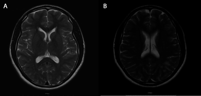
Figure 2.
Follow-up MRI images after 4 months show complete resolution of the lesion in the splenium of the corpus callosum on axial T2-weighted images (A). Extensive patchy T2 hyperintensities are noted in the deep and subcortical white matter (B) and likely to represent cerebral vascular disease in the context of illicit drug use.

