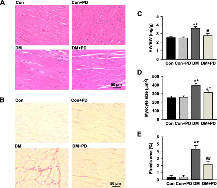
Fig. 3.
Polydatin attenuates diabetes-induced cardiac remolding. a Representative cross-sections of rat left ventricles histochemically stained with H&E staining. b Representative images of myocardium with cardiac fibrosis with Sirius red staining. c Heart weight-to-body weight ratio (HW/BW) of rats in different groups after 8-week polydatin or vehicle treatment. d Bar graph shows quantitative analysis of cardiomyocyte cross-sectional areas. e Bar graph shows quantified interstitial fibrotic areas (%). The scale in the images is 50 μm. Magnification × 200. Results were expressed as means ± SEM (n = 7). *P < 0.05, **P < 0.01 versus Con; #P < 0.05, ##P < 0.01versus DM

