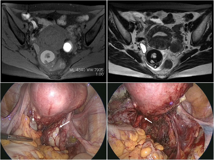
Fig. 1.
Magnetic resonance images show a right ovarian tumor with a mural nodule and a left ovarian endometrioma. a Transverse T1-weighted fat-saturated image after gadolinium enhancement. b Transverse T2-weighted image. c Laparoscopic findings before surgery of the bilateral ovarian mass with adnexal adhesion. White dotted arrow indicates left ovarian endometrioma, and white solid arrow indicates right ovarian tumor. d Laparoscopic findings after bilateral salpingo-oophorectomy. White arrow indicates lesions suspected as deep infiltrating endometriosis lesions in the left uterosacral ligament

