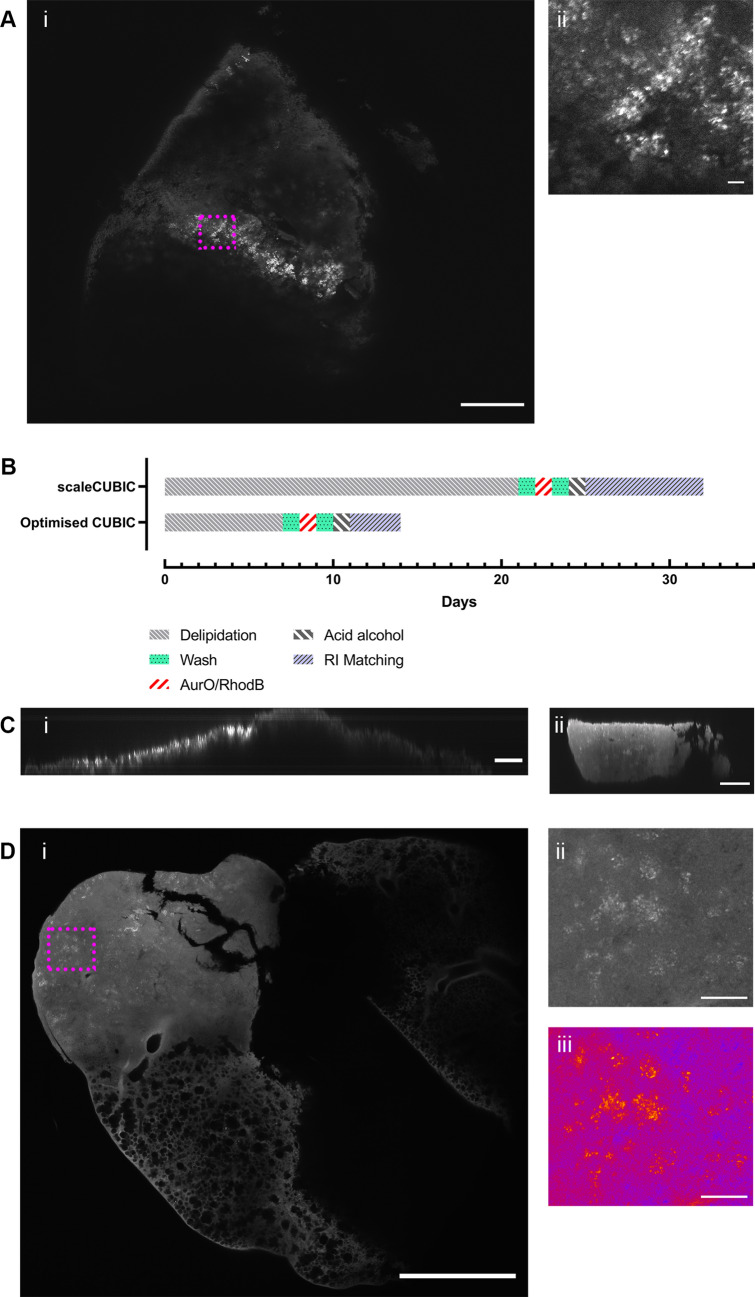
Figure 3.
Mesoscopy of Mtb infected lung. Mtb can be visualized using CAF staining procedure in GC1237 infected mouse lung slice (Ai) with a speckly pattern not seen in naïve controls or in tissue without lesions (supplementary Fig. 4) and is present in a zoomed-in image (Aii). However, using optimised CUBIC reagents30 processing time (B) can be reduced and the depth of imaging possible can be increased substantially, with scaleCUBIC (Ci) and comparable optimised CUBIC (Cii) presented. Using the optimised CAF procedure it is possible to see Mtb in a H37Rv infected sample (Di) and in zoomed-in images (Dii), with pseudo-colouring to highlight the speckled pattern (Diii). Scale bar = 500 µm (Ai, Di), 200 µm (C) and 20 µm (Aii, Dii, Diii).

