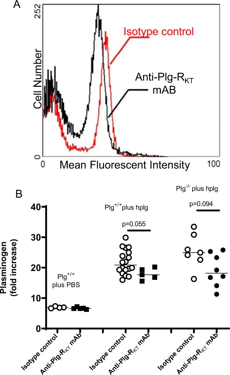
Fig. 2. Role of Plg-RKT in plasminogen accumulation in wounds.

A Effect of anti- Plg-RKT mAB on plasminogen binding to the surfaces of leukocytes. Plg+/+ and Plg−/− mice (8–12 weeks of age) were intravenously injected with 100 µl of anti-Plg-RKT mAB7H1 (2.5 mg/ml) or with mouse IgG2A (2.5 mg/ml) as isotype control (n = 3 for each study group). Thirty minutes later, standard burn wounds were introduced and all mice were intravenously injected with 100 µl (2 mg) of Alexa Fluor 488-labeled human plasminogen or PBS (as control). At 24 h after wounding and injection, blood samples were collected. Erythrocytes were lysed immediately with a solution containing 0.15 M NH4Cl for 5 min, and the remaining leukocytes were washed and resuspended in 500 μl PBS. FACS analysis was performed using a Cytomics FC500 (Beckman Coulter, Indiana, USA) and leukocytes were defined by forward scatter and side scatter. A clear peak shift of plasminogen binding to leukocytes was observed after anti-Plg-RKT mAB treatment, relative to injection with isotype control. B Effect of anti-Plg-RKT mAB on the accumulation of plasminogen in wounds. Both Plg−/− and Plg+/+ littermates were injected intravenously with isotype control or anti-Plg-RKT mAB (2.5 mg/ml) 30 min before the burn injury, followed immediately by intravenous injection of human plasminogen (hplg) (2 mg). Wound tissue was collected 24 h after the injury. The plasminogen concentration in wound lysates was determined by specific ELISA. Data analysis by robust ANOVA showed a significant effect of the addition of plasminogen (p < 0.001, f = 39.649), and a significant effect of antibody treatment (p = 0.003, f = 9.847), and a trend for antibody treatment × addition of plasminogen interaction (p = 0.084, f = 2.629). Post hoc testing by Mann–Whitney test showed: Plg-RKT+/+ plus hplg, isotype control (Mdn = 21), anti-Plg-RKT mAB (Mdn = 18), (U = 19, p = 0.055); Plg-RKT−/− plus hplg, isotype control (Mdn = 24.94), anti-Plg-RKT mAB (Mdn = 8.18), (U = 13, p = 0.094), n = 5–18.
