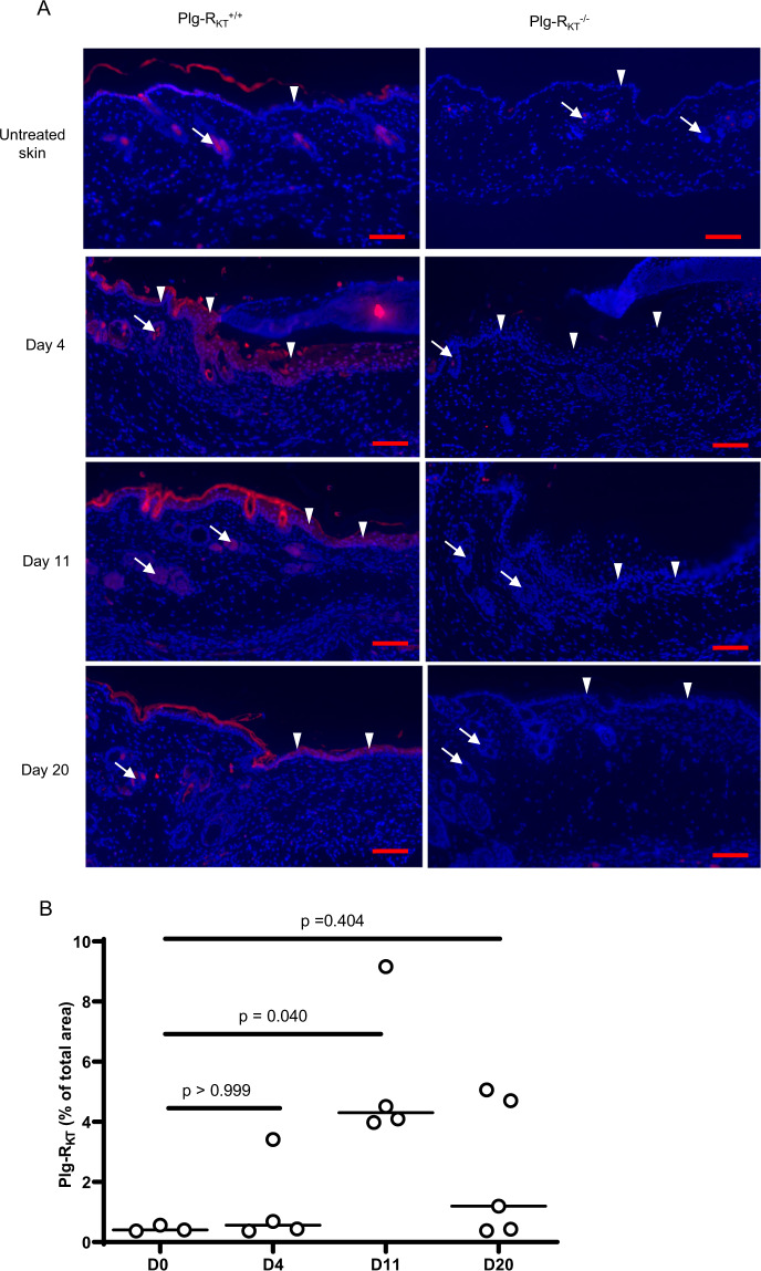
Fig. 3. Expression of Plg-RKT in wound tissue.
A Paraffin sections of wound tissue from Plg-RKT−/− and Plg-RKT+/+ littermates were immunostained with anti-Plg-RKT mAB (red) and DAPI (blue). Scale bar = 200 µm. White arrows indicate hair follicles, white arrowheads show keratinocytes. B Quantitation of the Plg-RKT staining in Plg-RKT+/+ (shown as % of total wound area). By Krustal–Wallis test, p = 0.054, Krustal–Wallis statistic = 7.1 followed by Dunns Multiple Comparisons, Day 0 vs. Day 4, p ≥ 0.999, Day 0 vs. Day 11 p = 0.040 and Day 0 vs. Day 20, p = 0.404, (n = 4). Medians are displayed. Images were captured with a Zeiss Axio Imager Z1 (Zeiss, Oberkochen, Germany) or Nikon A1R Eclipse Ti-E inverted microscope (Nikon Instruments, Amsterdam, Netherlands). Quantification of fluorescent areas was performed using Image J software.

