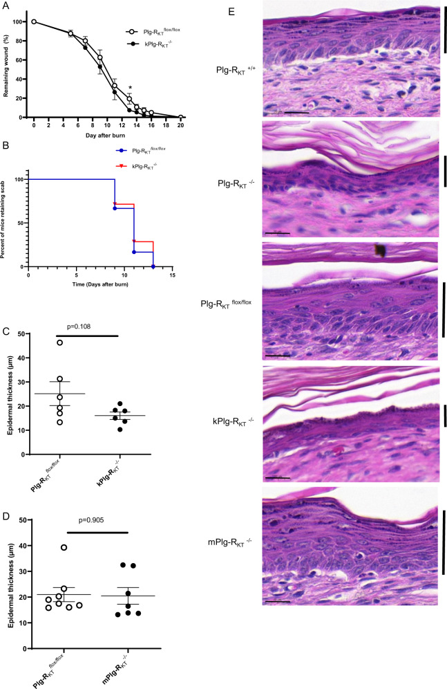
Fig. 5. Role of keratinocyte Plg-RKT in burn wound healing.
A Quantification of the remaining wound area at different time points after wounding of male kPlg-RKT−/− mice and Plg-RKT flox/flox mice (n ≥ 7). The mixed-effects model (REML), showed a significant effect for time (p < 0.0001, f = 211.9), and a trend for genotype (p = 0.103, f = 3.158) but no significant genotype × time interaction (p = 0.745), f = 0.658). Post hoc testing by two-tailed t test *p = 049. B Scab loss data are shown for cohorts of Plg-RKTflox/flox and kPlg-RKT−/− mice) (n = 7). p = 0.669, Χ2 = 0.182, df = 1 by the log-rank (Mantel–Cox) test. C Quantification of epidermal thickness at day 20 after wounding of kPlg-RKT−/− and Plg-RKTflox/flox mice. D Quantification of epidermal thickness at day 20 after wounding of mPlg-RKT−/− and Plg-RKTflox/flox mice. Means ± S.E.M. determined by two-tailed t test is shown in (A, C, D). E Representative images of H&E stained sections taken from the middle part of the healed area at day 20 after wounding from mice with different genotypes. Scale bar = 20 µm. The keratinocyte layer is marked with a vertical bar. Images were taken using a Leica DC300F digital camera attached to a Leica DM LB microscope (Leica, Wetzlar, Germany). Epidermal thickness was measured from the photos using Adobe Photoshop.

