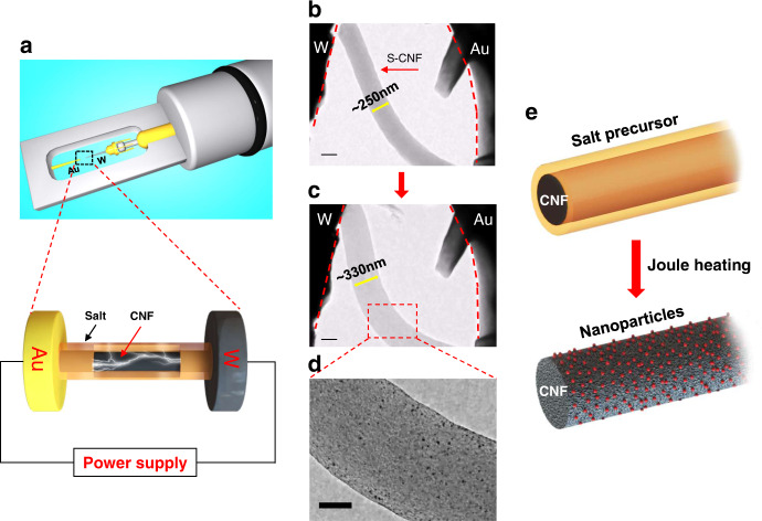
Fig. 1. In situ TEM observation of Pt nanoparticles formation on H2PtCl6-loaded CNF through electrical Joule heating.
a Schematic images of electrical biasing TEM holder and the zoomed-in view of the salt-loaded nanofibers between Au and W electrodes. b–d TEM images depict the CNF expansion and the formation of Pt nanoparticles on CNF. Scale bars are 200 nm in (b, c), 100 nm in (d). e The schematic of the S-CNF before and after the Joule heating.

