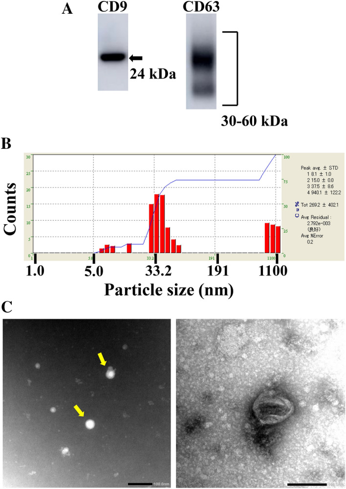
Figure 1.

Confirmation of isolation of exosomes from human serum, using western blot analyses of CD9 and CD63 levels (A), size distribution (B), and morphology (C). Human serum-derived exosomes were isolated using a kit. (A) The lysate of the resulting exosome fraction was resolved by SDS-PAGE and electroblotted onto a PVDF membrane. The blot was probed with anti CD9- and CD63-antibodies, followed by a corresponding HRP-tagged secondary antibody. Data are representative of three experiments. Full-length blots are present in Supplemental Fig. 1. (B) Isolated exosomes were suspended in 100 µL PBS. The size of the exosomes was measured by dynamic light scattering. (C) The morphology of exosomes was examined by TEM. Scale bar = 100 nm.
