
Figure 3.
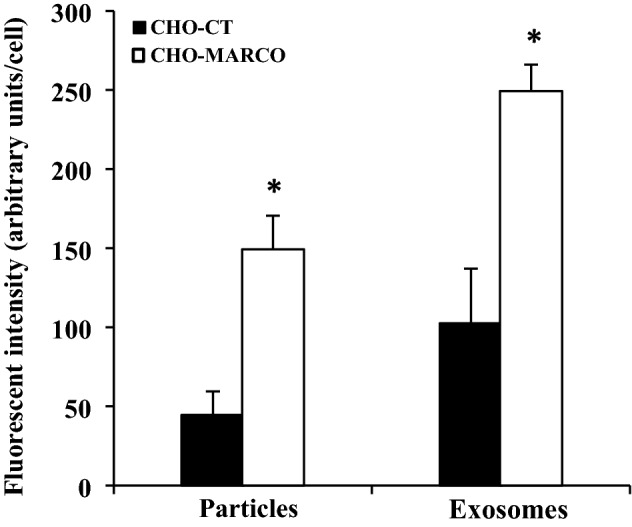
Quantification analyses of the association of exosomes or 20-nm nanoparticles with CHO-CT and CHO-MARCO cells. T-REx-CHO-MARCO cells were cultured in a 96-well plate with (CHO-MARCO) or without (CHO-CT) tetracycline. After 24 h of culture, the cells were treated with red fluorescent exosomes or 20-nm red fluorescent nanoparticles for 6 h. After incubation, the cells were washed twice with HBSS and fixed with 4% paraformaldehyde, followed by staining with DAPI and phalloidin. The associated fluorescent amounts were analyzed using an IN Cell Analyzer. Data are presented as means ± SEM (n=3). *, Significantly different from CHO-CT cells.