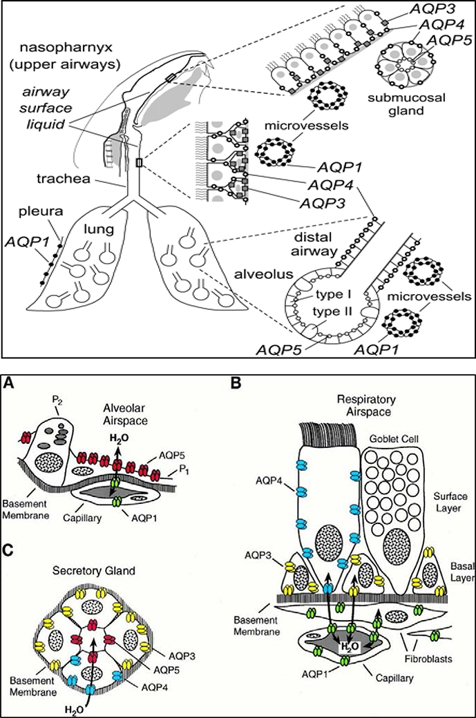
FIGURE 3: AQP expression in airways and lung compartments.
Upper Panel: Schematic diagram showing that mouse AQPs 1, 3, 4 and 5 are expressed in epithelia and endothelia throughout the nasopharyngeal cavity, airways (upper and lower), and alveolar compartment.
Lower Panel: Schematic diagrams showing cellular expression patterns of AQPs in rat airways and lung tissues: A). Subcellular expression pattern of AQP5 in alveolar space. AQP5 (shown in red) is expressed in apical membrane of type 1 pneumocytes (P1) but not expressed in type 2 pneumocytes (P2) and AQP1 is expressed in the alveolar capillaries; B). Cellular expression pattern of AQPs in respiratory epithelial layer, depicting no expression in goblet cells, AQP3 expression in basal cells, AQP4 expression in surface columnar cells, and AQP1 in underlying capillaries and fibroblasts; C). Subcellular expression pattern of AQPs in secretory glands: AQP5 (red) in apical membrane of acinar cells, AQP3 (yellow) and 4 (blue) in basolateral domains of glandular cells of nasopharynx and conchus but not in salivary glands. Reproduced with permission the Upper panel image from Borok and Verkman 2002 [2] and the Lower panel images A, B, and C from Nielsen et al 1997 [10].

