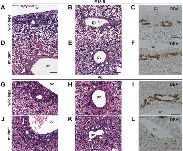
Figure 2.

Anks6 deficiency causes bile duct abnormalities. (A, B, D and E) Hematoxylin and eosin-stained liver sections of E18.5 wild type (A and B) and Anks6 mutant (D and E) embryos. In wild-type liver, bile ducts are lined by cuboidal epithelium and surrounded by portal mesenchyme in the hilar region (A). Less differentiated bile ducts are observed in the peripheral area (B). In the Anks6−/− liver, note the presence of multiple, low-cuboidal bile ducts in the hilar (D), as well as in the peripheral (E) area, indicative of a failure in ductal plate remodeling. Green arrows indicate bile ducts; pv, portal vein. Scale bar, 50 μm. (G, H, J and K) Hematoxylin and eosin-stained liver sections of neonatal wild type (G and H) and mutant (J and K) mice. Both hilar and peripheral bile ducts are embedded in the periportal mesenchyme in wild-type liver at this stage (G and H). In contrast, the bile ducts remain in a ductal plate conformation in the mutant livers (J and K). Green arrows indicate bile ducts. Scale bar, 50 μm. (C, F, I and L) DBA expression in E18.5 (C and F) and neonatal (I and L) liver. DBA is expressed in the entire biliary epithelium in wild-type livers at E18.5 (C) and P0 (I). In the mutant liver, DBA expression is limited to the portal side of the bile duct at E18.5 (F) and has scattered biliary expression at P0 (L), indicative of a failure in biliary epithelium differentiation in the mutant liver. Scale bar, 50 μm.
