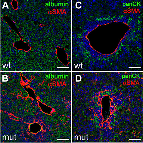
Figure 3.

Loss of Anks6 causes portal fibrosis. (A–D) Immunofluorescent analysis of αSMA expression in E18.5 wild type (A and C) and Anks6 mutant (B and D) livers. αSMA is expressed in a layer of perivascular cells underneath the portal endothelium in wild-type livers (A and C). In Anks6 mutant livers, αSMA expression extends throughout the periportal mesenchyme and αSMA-positive cells can be seen in the albumin-positive (green) liver parenchyma (B), and they envelope the pan-cytokeratin-positive bile ducts (panCK, green) (D), indicating a conversion of the periportal mesenchyme to myofibroblasts. Scale bar, 75 μm.
