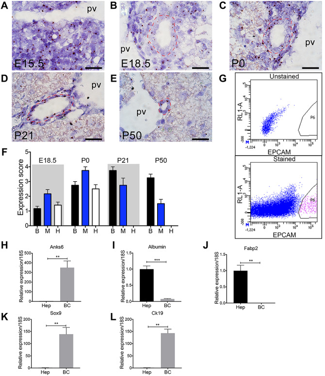
Figure 4.

Anks6 is expressed in the biliary epithelium and portal mesenchyme. (A–E) Anks6 expression analysis using in situ hybridization in the liver at E15.5 (A), E18.5 (B), P0 (C), P21 (D), and P50 (E). Representative images of Anks6 staining reveal its expression in the hepatoblast progenitor cells at E15.5 (A), portal mesenchyme, bile duct epithelium and hepatocytes around birth (B and C), and in the biliary epithelium and surrounding mesenchyme at adult stage (D and E). Red dashed line surrounds bile ducts. pv, portal vein. Scale bar, 22.5 μm. (F) Anks6 expression scoring in wild-type liver during development and postnatal stages (0–4 arbitrary units). See representative images in (A). B, biliary cells; H, hepatocytes; M, portal mesenchyme. (G) Gating strategy for isolation of EpCAM+ biliary cells from the livers of 6-week old wild-type mice. Total single-cell suspensions of hepatocyte-depleted biliary and non-parenchymal cell fraction were sorted from the P6 population of EpCAM-stained samples. (H–L) Anks6 is expressed in biliary cells of adult liver. Hepatocytes (Hep) and biliary cells (bc) were isolated and relative mRNA expression of noted genes was quantitated via qRT-PCR. Hepatocytes are normalized to one and bc expression is quantitated as a ratio to its expression. Data represent the average of 3 biological samples ± s.e.m. Two-tailed unpaired t-test was used for statistical analysis, *P < 0.05, **P < 0.01, ***P < 0.001.
