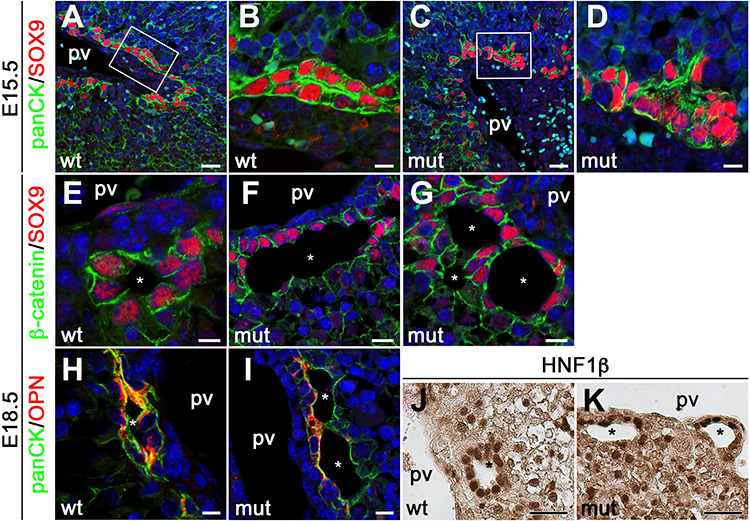
Figure 5.

Anks6 is required for bile duct morphogenesis and cholangiocyte differentiation. (A–D) Immunofluorescent analysis of developing bile ducts in wild type (A and B), and mutant (C and D), livers with antibodies against panCK and Sox9 at E15.5. Scale bar, (A and C) 25 μm; (B and D) 7.5 μm. (E–G) Immunofluorescent analysis of E18.5 bile ducts with antibodies against β-catenin and Sox9. Sox9 is expressed in the entire epithelium of differentiated bile ducts in wild-type livers (E). Only the portal side of the ducts (first ductal layer) is positive for Sox9 in mutant livers (F and G), indicating that cholangiocyte differentiation is impaired in Anks6 mutant livers. In addition to biliary differentiation defects the mutant bile ducts demonstrate abnormal lumen formation (G). pv, portal vein, white asterisk indicates bile duct lumen. Scale bar, (E) 7.5 μm; (F and G) 10 μm. (H and I) Antibody staining against the apical marker osteopontin, demonstrates normal apical polarization of E18.5 wild-type biliary epithelial cells (H). In mutant bile ducts, osteopontin is present in the portal side, but missing from the parenchymal side of the ducts (I). pv, portal vein, white asterisk indicates bile duct lumen. Scale bar, (H) 7.5 μm; (I) 10 μm. (J and K) Immunohistochemistry against the cholangiocyte differentiation marker Hnf1β. Hnf1β is present in both layers of the bile duct in wild-type livers (J), but restricted to the portal layer in mutant livers (K). pv, portal vein, black asterisk indicates bile duct lumen. Scale bar, 30 μm.
