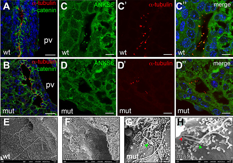
Figure 6.

Loss of Anks6 causes structural abnormalities in bile duct cilia. (A and B) Immunofluorescent staining of wild type (A) and Anks6 mutant (B) bile ducts with acetylated α-tubulin (red, cilia) and β-catenin (green, bile ducts) demonstrates the lack of cilia from the parenchymal side of the bile duct in the mutant liver at E18.5. Scale bar, 25 μm. (C–D″) Anks6 localizes to cholangiocyte cilia in E18.5 livers. Antibody staining against Anks6 (green) and acetylated α-tubulin (red, cilia) shows Anks6 ciliary localization in wild-type cholangiocyte cilia (C, C′ and C″), and its absence from the mutant cilia (D, D′ and D″). Scale bar, 10 μm. (E–H) Scanning electron microscopy images reveal ciliary abnormalities in Anks6 mutant livers. Wild-type cilia are long and solitary (E), whereas mutant cilia are short (F), with bulbous structures (green arrowhead) (G), or are duplicated at the basal body (red arrowhead), and appear kinked (green arrowhead) (H). Scale bar, (e, F and G) 1 μm, (h) 100 nm.
