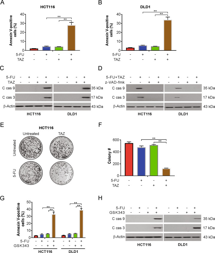
Fig. 2. Tazemetostat enhanced 5-FU-mediated apoptosis in CRC cells.
A, B HCT116 cells were treated with 2.5 μg/mL 5-FU, 0.5 μM tazemetostat, or their combination for 24 h. Apoptosis was analyzed by flow cytometry. C HCT116 or DLD1 cells were treated with 2.5 μg/mL 5-FU, 0.5 μM tazemetostat, or their combination for 24 h. Indicated proteins were analyzed by western blotting. D HCT116 or DLD1 cells were treated with the combination of 2.5 μg/mL 5-FU and 0.5 μM tazemetostat with or without z-VAD-fmk pretreatment for 24 h. Indicated proteins were analyzed by western blotting. E, F HCT116 cells were treated with 2.5 μg/mL 5-FU, 0.5 μM tazemetostat, or their combination for 2 h. After 2 weeks, the plates were stained for cell colonies with crystal violet dye. G HCT116 or DLD1 cells were treated with 2.5 μg/mL 5-FU, 1 μM GSK343, or their combination for 24 h. Apoptosis was analyzed by flow cytometry. H HCT116 or DLD1 cells were treated with 2.5 μg/mL 5-FU, 1 μM GSK343, or their combination for 24 h. Indicated proteins were analyzed by western blotting. The results were expressed as the mean ± SD of three independent experiments. **P < 0.01.

