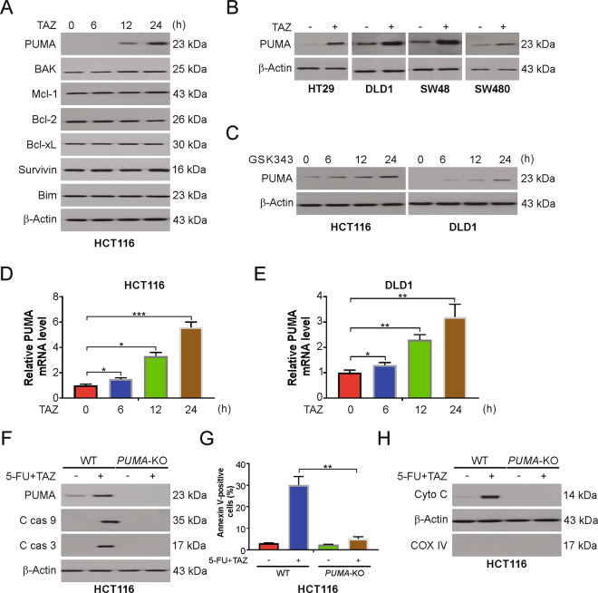
Fig. 3. PUMA is required for tazemetostat/5-FU-induced apoptosis.
A HCT116 cells were treated with 0.5 μM tazemetostat at indicated time points. Indicated proteins were analyzed by western blotting. B Indicated cell lines were treated with 0.5 μM tazemetostat for 24 h. PUMA expression was analyzed by western blotting. C HCT116 or DLD1 cells were treated with 1 μM GSK343 at indicated time points. PUMA expression was analyzed by western blotting. D HCT116 cells were treated with 0.5 μM tazemetostat at indicated time points. mRNA level of PUMA was analyzed by real-time PCR. E DLD1 cells were treated with 0.5 μM tazemetostat at indicated time points. mRNA level of PUMA was analyzed by real-time PCR. F WT and PUMA-KO HCT116 cells were treated with the combination of 2.5 μg/mL 5-FU and 0.5 μM tazemetostat for 24 h. Indicated proteins were analyzed by western blotting. G WT and PUMA-KO HCT116 cells were treated with the combination of 2.5 μg/mL 5-FU and 0.5 μM tazemetostat for 24 h. Apoptosis was analyzed by flow cytometry. H Cytosolic fractions isolated from WT and PUMA-KO HCT116 cells treated with the combination of 2.5 μg/mL 5-FU and 0.5 μM tazemetostat for 24 h were probed for cytochrome c by western blotting. β-actin and cytochrome oxidase subunit IV (Cox IV), which are expressed in cytoplasm and mitochondria, respectively, were analyzed as the control for loading and fractionation. The results were expressed as the mean ± SD of three independent experiments. *P < 0.05; **P < 0.01.

