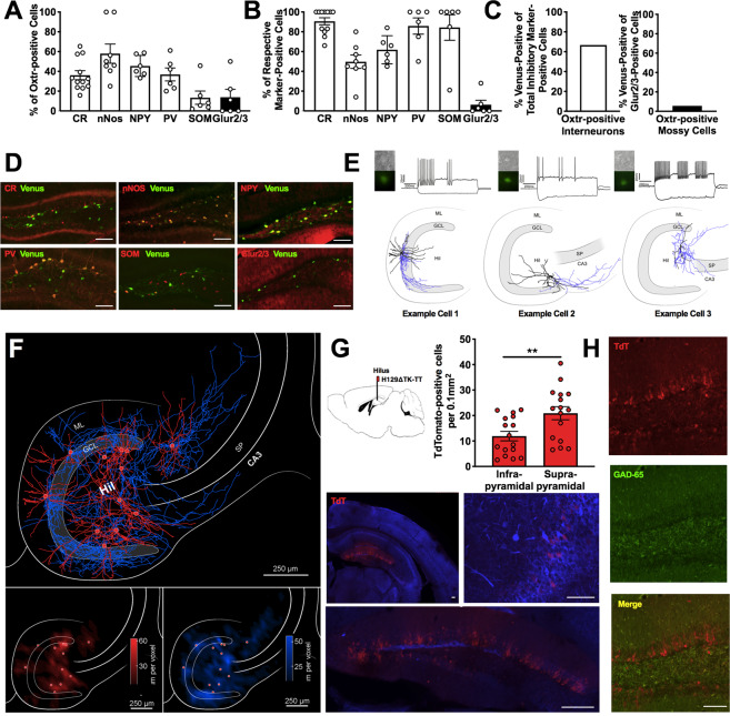
Fig. 2. Oxtr-positive hilar neurons are a diverse population of interneurons which project to DGGC.
a Quantification of Oxtr colocalization with respective marker, expressed as a percentage of total Oxtr-positive cells. n = 6CR/4nNOS/3NPY/3PV/3SOM/3Glur2/3 mice per group. b Quantification of Oxtr colocalization with respective marker, expressed as a percentage of marker-positive cells n = 3–6 mice per group. c Quantification of Oxtr colocalization with total interneuron markers or mossy cell marker, expressed as a percentage of total interneuron marker-positive or Glur2/3-positive, respectively. d Representative images of Oxtr and respective markers of interneurons and mossy cell coexpression in the DG. e Oxtr-positive hilar neurons were identified visually and recorded in voltage-clamp configuration mode. Heterogeneous firing patterns were observed. f Summary plot of all reconstructed axonal (blue) and dendritic (red) distribution patterns of Oxtr-HI merged into a map of the hippocampus. Note axonal arborization covering the entire DG. Also notice the presence of individual collaterals exiting the DG and projecting to area CA3 of the hippocampal formation. Bottom inset, quantitative analysis of the dendritic, bottom left, and axonal, bottom right, domains, as shown in contour plot indicating the density of either the dendritic and axonal segments with respect to their position in the hippocampus. Note that the dendritic domains are mainly restricted to the layer of the origin of somata (red dots). Oxtr-HI axons show a distributed density pattern with projections into the dentate granule cell layer, stratum moleculare, and CA3 stratum oriens, stratum lucidum, stratum radiatum, and stratum lacunosum moleculare. Voxel size 50 μm3. g Representative widefield florescent microscopy images showing TdT-positive cell bodies following injection of Cre-dependent transsynaptic anterograde tracer in the hilus of Oxtr-Cre mice. Note the presence of TdT-positive cell bodies in the DG GCL, and modest expression in the hilus and CA3 stratum pyramidale. Quantification of TdT-positive neurons in the suprapyramidal and infrapyramidal DG blades, respectively. A difference was observed, with significantly more TdT-positive cell bodies in the suprapyramidal blade. n = 8 mice; t = 2.836, df = 30, P = 0.0081. h TdT-positive cell bodies in the GCL were rarely colocalized with GAD65 positive neurons. Scale bars: 250 μm. **P < 0.01. ML moleculare layer, GCL granule cell layer, Hil Hilus, SP stratum pyramidale.

