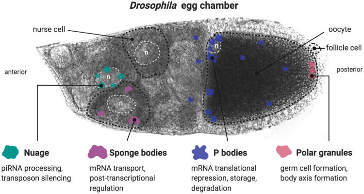
Figure 1. Schematic and role of maternal granules in egg chambers.
Nuage is localised around the nurse cell nuclei, while sponge bodies are dispersed throughout the cytoplasm of the nurse cells. P bodies are enriched at the anterior margin of the oocyte (especially in the dorso-anterior corner). They are also observed throughout the oocyte and nurse cell cytoplasm. Polar granules are present at the posterior pole of the oocyte. Fifteen nurse cells, positioned to the anterior, produce the components (mRNAs, proteins, etc.) required for the development of a single oocyte. These germline-derived cells are interconnected through cytoskeletal bridges, allowing for cytoplasmic movement between them, and are surrounded by somatic-derived layer of follicle cells. (Representative cell types of the egg chamber are outlined with black dotted lines. Representative nuclei are outlined with white dotted lines and marked with an ‘n’). Created in BioRender.

