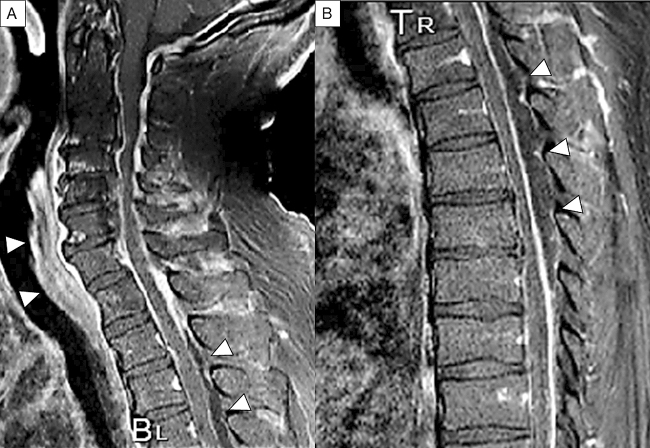
Figure 2.
Sagittal Gd-enhanced T1-weighted MRI. Spinal epidural abscess is noted that a capsule-like rim whose edges are strongly enhanced and the content of abscess shows low intensity in the cervical (A) and thoracic (B) spine. The retro-pharyngeal abscess is also noted as enhanced mass (A). Arrowheads indicate the abscess.

