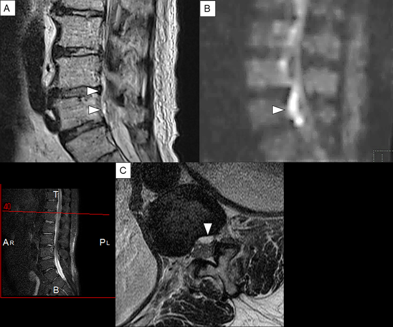
Figure 5.
Sagittal T2-weighted and Gd-enhanced MRI of the lumbar spine in case2.
(A): Sagittal view of T2-weighted MRI.(B): Sagittal view of diffusion-weighted image. (C): Axial view of T2-weighted MRI. A linear lesion that shows high intensity and appears to be an epidural abscess that is found on the dorsal side of the vertebral body (A). The same lesion shows a strong high signal area in the diffusion-weighted image (B). This finding is considered to reflect the decrease in diffusion of water molecules in the abscess lumen and is characteristic findings of abscess. The axial view confirms anterior SEA that shows high signal intensity on the dorsal side of the vertebral body at the level of L1 (C). Arrowheads indicate SEA.

