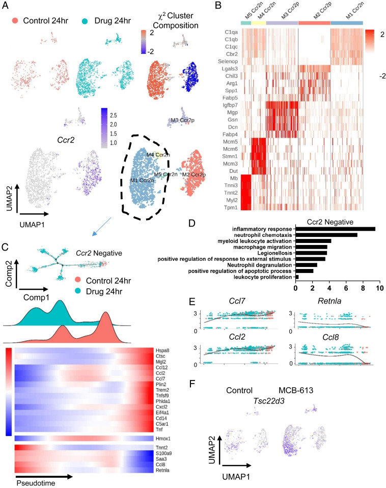
Fig. 4.
Single cell analysis of macrophages 24 h post-MI reveals that the MCB-613 protective response is associated with increased anti-inflammatory macrophages and macrophage and granulocyte functions. (A) UMAP plot of macrophages from control (salmon) and MCB-613 (aqua) 24 h post-MI. The χ2 cluster composition analysis of subclustered macrophages from MCB-613 compared to control at 24 h post-MI (Top Right). UMAP plot of macrophage subclusters 24 h post-MI (Bottom Right). (B) Heat map showing top five differentially expressed genes. (C) Pseudotime trajectory and density plot of Ccr2n macrophages across the pseudotime trajectory. Heatmap showing dynamics of gene expression for Ccr2n macrophages across the trajectory. (D) GO terms analysis of differentially expressed genes from macrophages in control compared to MCB-613. (E) Gene expression trends for individual genes across the pseudotime trajectory. (F) Feature plot showing Tsc22d3 expression in macrophages for control and MCB-613.

