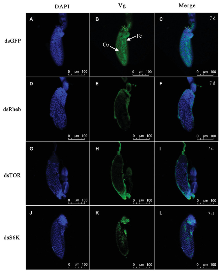
Figure 7.
Silencing of TOR pathway-related gene changes Vg accumulation in ovarioles of adult females at 7 DAE. Left panels (A,D,G,J) show DAPI-stained nuclei; middle panels (B,E,H,K) show Vg protein detected with goat anti-rabbit IgG-labeled with Dylight 488 (green); right panels (C,F,I,L) showed merged images. Fluorescent images were captured with a Zeiss LSM 780 confocal microscope (Carl Zeiss MicroImaging, Göttingen, Germany). Fc, follicular cell; Oo, oocyte. Bars, 100 μm.

