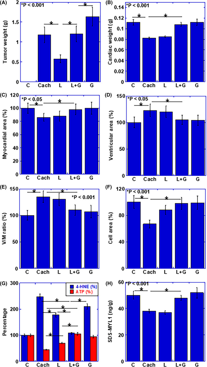
Figure 5.

Alteration of myocardium and effect of LAA and glucose in mouse cachectic model. A, Weight of peritoneal tumor. B–H, Effect of LAA and glucose in cachectic mice on B, cardiac weight, C, myocardial area, D, ventricular area, E, ventricular area to myocardial ratio (V/M ratio), F, mean cardiomyocyte area (cell area), G, concentrations of ATP and 4‐HNE in myocardium, and H, protein level of SDS‐soluble myosin light chain (SDS‐MYL1). Error bars, standard error from 3 mice. Statistical significance was calculated by Student t test. C, no tumor control; Cach, cachexia mice; HNE, hydroxynonenal; G, glucose; SDS,‐MYL1, L, lauric acid; sodium dodecyl sulfate‐soluble myosin light chain‐1
