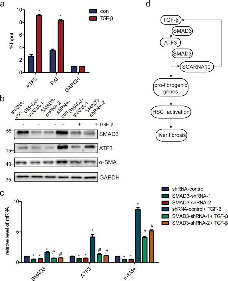
Fig. 7. ATF3 is over-expressed in a TGF-β/SMAD3-dependent manner.
a LX-2 cells were treated with TGF-β and ChIP analyses were performed on ATF3 promoter regions, using anti-SMAD3 antibody. Enrichment was shown relative to input; *p < 0.05. b, c LX-2 cells were infected with lentivirus-mediated shSMAD3-1/2 for 72 h and further treated with 10 ng/ml TGF-β for additional 24 h. The level of SMAD3, ATF3, and α-SMA were detected by western blot and qRT-PCR. d Schematic representation of the TGFβ/ATF3/lnc-SCARNA10 pathway and its function in the progression of liver fibrosis. The data were expressed as the mean ± SEM for at least triplicate experiments. GAPDH was used as an internal control; */#p < 0.05, *p < 0.05 for vs shRNA control, # p < 0.05 for vs shRNA control + TGF-β.

