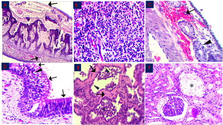
Figure 2.
Histopathological changes in the MERS-CoV natural infected dromedary camels in Saudi Arabia 2018–2019.
A: Nasal Turbinate showed exfoliation of the epithelial cells leaving denuded basement membrane (arrow), x100; B: Nasal Turbinate showed sub-epithelial mononuclear inflammatory cells infiltration (arrow), x400; C: Nasal Turbinate showed sub-epithelial hemorrhage (arrow) and glandular degeneration (arrowhead), x400; D: Trachea showed partial loss of cilia cells (arrow) and epithelial vacuolation (arrowhead), x400; E: Lung showed marked hyalinization of alveolar septa (arrow), with hypertrophy of pneumocytes type II (arrowhead) and intra-alveolar accumulation of macrophages (asterisk), x400; F: Renal Corpuscle showed marked fibrinous exudate with complete damage of glomerular tuft (asterisk), x400; all slides were stained with H&E stain.

