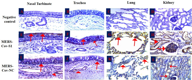
Figure 4.
Immunohistochemistry of the MERS-CoV natural infected dromedary camels. A-D: Nasal turbinate, trachea, lungs and kidney respectively showed no signal; E-F: Nasal turbinate and trachea showed signals of MERS-CoV-S1 viral antigen in the epithelial surfaces; G: pulmonary alveoli showed signals of MERS-CoV-S1 viral antigen, H: MERS-CoV-S1 antigen was detected in both glomerular epithelia (arrow) and renal tubules (arrow head); I-J: Nasal turbinate and trachea showed weak signals of MERS-CoV-NC viral antigen in both epithelial surfaces (arrow) and sub-epithelial glands (arrowhead); K: Pulmonary alveoli showed weak signals of MERS-CoV-NC viral antigen; L: Renal tubules showed weak signals of MERS-CoV-NC viral antigen.

