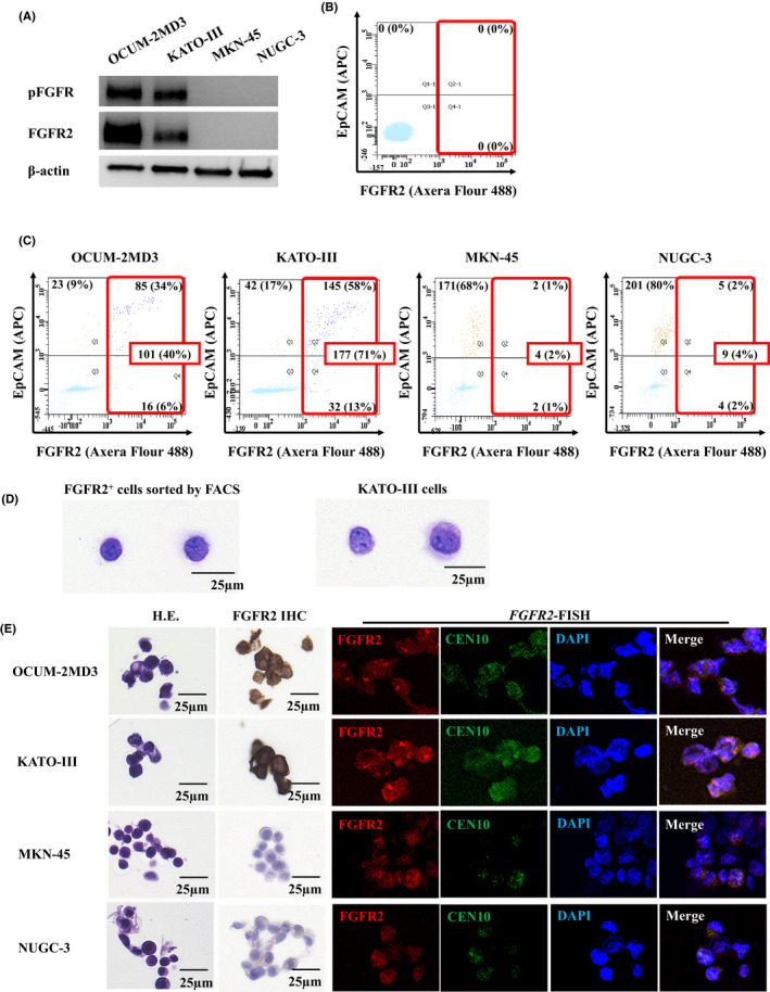
Figure 1.

Fibroblast growth factor receptor2 (FGFR2) expression and determination of gastric cancer cells in peripheral blood. A, Western blot analysis of FGFR2/phospho‐FGFR (pFGFR) expression. KATO‐III cells and OCUM‐2MD3 cells expressed both FGFR2 and pFGFR2, but MKN45 cells and NUGC3 cells did not. B, Determination of FGFR2+ cells in peripheral blood of a healthy volunteer by flowcytometry. No FGFR2+ cell was detected when the cutoff value of FGFR2+ fluorescence was 1000. C, Detection of FGFR2+ gastric cancer cells in the peripheral blood of a healthy volunteer by flowcytometry. FGFR2+ cells of OCUM‐2MD3, KATO‐III, MKN45, and NUGC3 were detected (101 cells, 177 cells, 4 cells, and 9 cells of a total of 250 cells, respectively). D, H&E staining of FGFR2+ cells and cultured KATO‐III cells. The morphology of FGFR2+ cells sorted by FACScan is similar to that of cultured KATO‐III cells. E, FGFR2 immunostaining and FGFR2 FISH of cancer cell lines sorted by FACScan. FGFR2 expression and FGFR2 amplification were found in OCUM‐2MD3– or KATO‐III–spiked peripheral blood but not in MKN45– or NUGC–spiked peripheral blood
