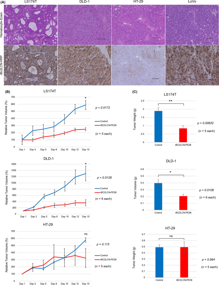
FIGURE 2.

Evaluation of the therapeutic efficacy of rBC2LCN‐PE38 in cell line‐derived mouse xenograft models in vivo. (A) Findings of histochemical staining for each cell line‐derived subcutaneous tumor collected from mouse xenograft models (scale bar: 100 µm). Histological differentiation/rBC2LCN expression were: LS174T, well‐to‐moderate/strong; DLD‐1, poor/strong; HT‐29, poor/weak; and LoVo, poor/strong. (B) Change of relative tumor volume during the experimental period. Tumor size was measured in two dimensions by digital calipers, and the volume was calculated using the following formula: 0.5 × width2 × length. The tumor volume on Day 1 was defined as the standard volume (100%). (C) Excised tumor weight from mouse xenograft models. *P < 0.05, **P < 0.01. ns, not significant
