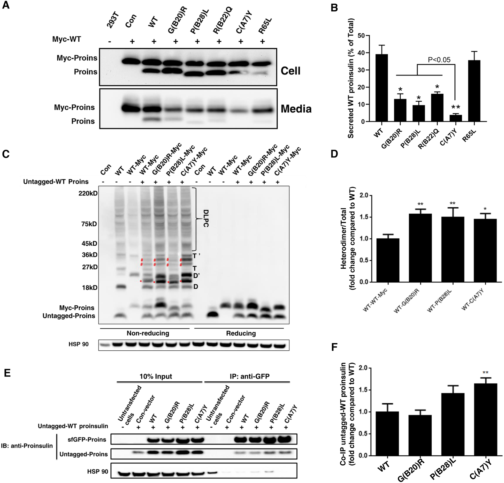
Fig.4. Proinsulin mutants interact with co-expressed WT-proinsulin and impair the ER export of WT-proinsulin.

A-B. 293T cells were co-transfected with Myc-tagged WT-proinsulin (upper bands) and untagged WT-proinsulin or mutants (lower bands) as indicated. The secretion of Myc-tagged WT-proinsulin in the presence of untagged WT-proinsulin or mutants under 24h steady state was examined by immuno-blotting using anti-proinsulin. The percentages of secreted WT-proinsulin were quantified and calculated. * p <0.05 and ** p <0.01 comparing to WT (ANOVA test). C-D. 293T cells were co-transfected with untagged WT-proinsulin and Myc-tagged WT-proinsulin or mutants. The monomers, dimers (D refers to homodimers formed by untagged Proins, and D’ refers to homodimers formed by Myc-Proins, red star refers to heterodimers formed by untagged Proins and Myc-Proins), trimers (T refers to homotrimers formed by untagged Proins, and T’ refers to homotrimers formed by Myc-Proins, blue star refers to heterotrimers formed by untagged Proins and Myc-Proins), and higher-molecular weight disulfide-linked proinsulin complexes (DLPC) were analyzed under non reducing conditions. The total amount of untagged WT-proinsulin and Myc-tagged WT or mutants were analyzed under reducing condition. The percentages of heterodimer (red star marked) formed by untagged WT Proins and Myc-tagged proinsulin mutant were calculated. The percentage of heterodimer formed by untagged Proins-WT and Myc tagged Proins-WT was set to 1. * p <0.05 and ** p <0.01 comparing to WT (ANOVA test). E-F. 293T cells were co-transfected with untagged WT-proinsulin and super folder (sf) GFP-tagged WT-proinsulin or mutants. At 48 h post-transfection, cells were lysed and immunoprecipitated with the anti-GFP antibody, followed by immuno-blotting (IB) with anti-proinsulin antibody. The percentages of untagged WT-proinsulin pulled down by sfGFP-tagged proinsulin-WT or mutants were quantified and calculated. ** p <0.01 comparing to WT (ANOVA test).
