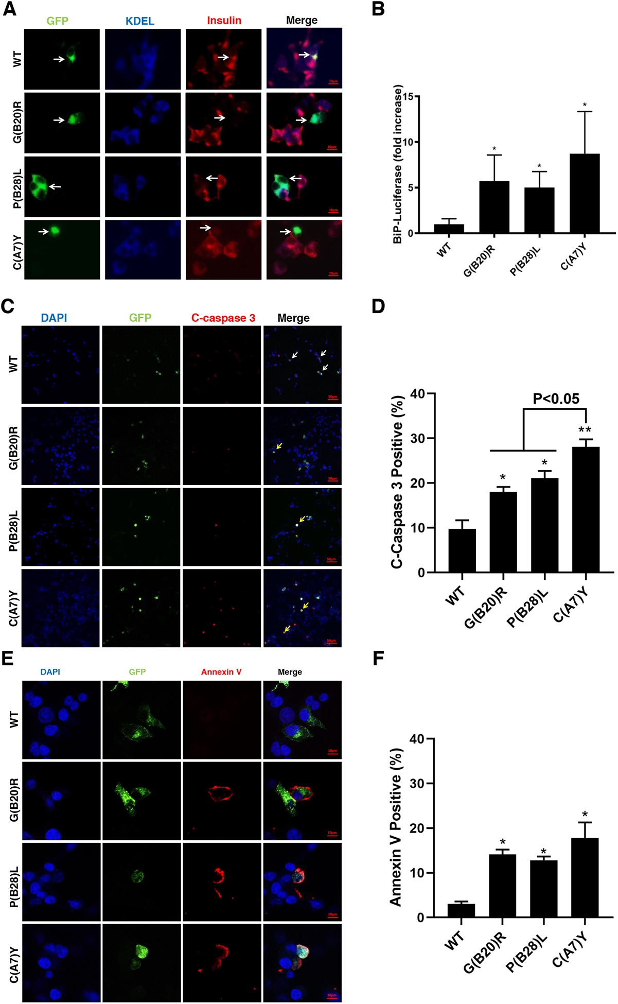
Fig.5. Proinsulin mutants decrease endogenous insulin production and induce ER stress, leading to apoptosis in beta cells.

A. INS1 cells were transfected with plasmid encoding sfGFP-tagged WT, G(B20)R, P(B28)L or C(A7)Y proinsulin. At 48h post-transfection, the cells were permeabilized and immunoblotted with anti-insulin (red) and anti-KDEL (blue, ER marker). Arrows indicate the cells expressed exogenous sfGFP-tagged WT-proinsulin or mutants.
B. INS1 cells were transiently triple-transfected with the plasmids encoding BiP promoter-firefly luciferase, CMV-driven Renilla luciferase, and WT or mutant proinsulin at ratio 1 : 2 : 5 (This ratio helps ensure that BiP-luciferase serves as a reporter from cells synthesizing exogenously expressed proinsulins). At 48 h post-transfection, the cells were lysed and a ratio of firefly/renilla luciferase was measured. The relative activities of the BiP promoter in cells expressing proinsulin mutants were compared to that in cells expressing WT-proinsulin, which served as a control and set to 1. Results are from at least three independent experiments. *p <0.05 compared with WT-proinsulin (Student’s t-test). C. INS1 cells were transfected with sfGFP-tagged WT-proinsulin and variants as indicated. After 3 days post transfection, cells were fixed and stained with anti-cleaved caspase 3 antibody. Arrows indicate cells expressed exogenous proinsulin with (yellow arrow) or without (white arrow) apoptosis. D. Percentages of cleaved caspase 3 positive cell in transfected INS1 cells were quantified. * p <0.05 and ** p <0.01 comparing to WT (Student’s t-test). E. Representative images of INS1 cells expressing sfGFP-tagged proinsulin stained with Annexin V (red) and DAPI (blue) were shown. F. Proportion of Annexin V positive cells were quantitative analyzed. *p <0.05 compared with WT-proinsulin (Student’s t-test)
