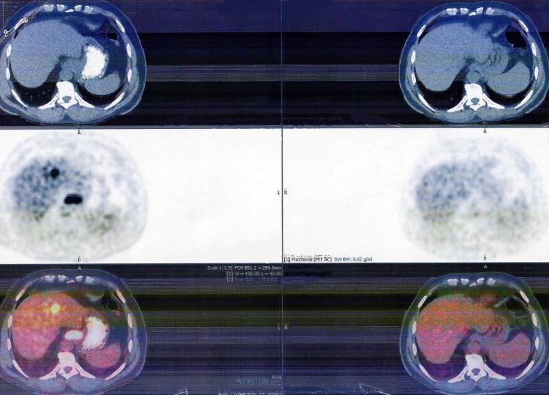
Figure 1. Positron Emission Tomography scan.
Towards the left side of the image is the initial scan at the time of diagnosis in August 2017, showing 18F-fluorodeoxyglucose avid lesions in the liver (metastatic lesions) and at the gastroesophageal junction. To the right is the last scan done in July 2020, which cannot show active metastatic disease process in response to trastuzumab monotherapy.

