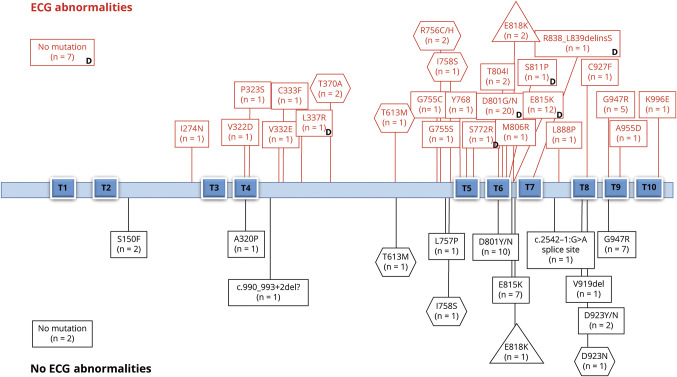
Figure 3. ECG abnormalities in ATP1A3-related syndromes.
Graphic representation of the ATP1A3-related syndromes (alternating hemiplegia of childhood [AHC] = rectangles, cerebellar ataxia, areflexia, pes cavus, optic atrophy, and sensorineural hearing loss [CAPOS] = triangles, rapid-onset dystonia-parkinsonism [RPD] = hexagons) (n = 110), associated mutations, and prevalence of ECG (12-lead and/or Holter) abnormalities. Each box includes the number of cases with a specific mutation and ECG abnormalities in the upper part or without ECG abnormalities in the lower part. Reference sequences for the corresponding ATP1A3 transcript and protein were NM_152296.4 and Uniprot P13637, respectively. T1 through T10 are transmembrane domains. The position of the mutation across the functional domains was not associated with different prevalence of ECG abnormalities or dynamic changes. In 9 patients with AHC, no mutation was identified in ATP1A3. D = dynamic changes (when serial tests were available).

