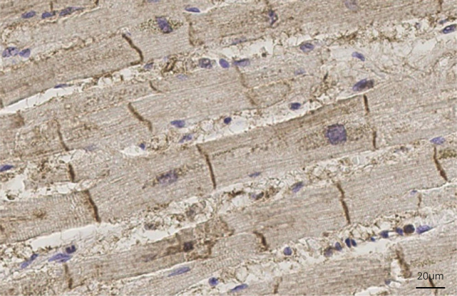
Figure 4. ATP1A3 expression in an adult normal heart.
Dark brown stripes show strong immunolabeling for ATP1A3 corresponding to intercalated disks in adult myocardium from a 75-year-old man (cause of death at postmortem: bronchopneumonia). Tissue samples were fixed in formalin and embedded in paraffin. A standard immunohistochemistry method was applied to 5-μm-thick sections with primary antibody anti-ATP1A3 (Santa Cruz, polyclonal, goat, sc16052) at a dilution of 1:1,000 with overnight incubation at 4°C in diluent buffer (DAKO REAL, Ab diluent S2022). Immunostaining was qualitatively evaluated.

