Fig. 6.
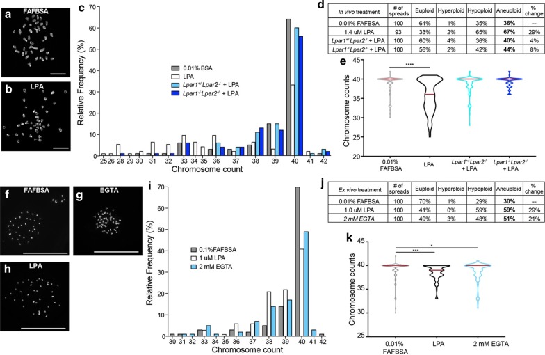
LPA signaling and AJ disruption enhances aneuploidy. a–e Chromosomal content from metaphase cells from E13.5 cortices injected with 0.01% FAFBSA (gray), 1.4 µM LPA (white), Lpar1+/−Lpar2−/− + LPA (pale blue), and Lpar1−/−Lpar2−/− + LPA (blue), assessed 6 h after injection a, b, Representative images of metaphase chromosomes stained with DAPI after in vivo exposure to (a) 0.01% FAFBSA (control) or b 1.4 μM LPA. Scale bar 10 μm. c Frequency of chromosome content in metaphase cells from E13.5 cortices assessed 6 h after injection. d Table of total counts and percentages; 100 metaphase spreads were counted for each treatment group. Euploidy = 40 chromosomes, Aneuploidy ≭ 40 chromosomes, Hyperploidy > 40 chromosomes and Hypoploidy < 40 chromosomes. e Violin plots of chromosome counts, assessed 6 h after injection. Red line denotes the median. Significance determined by nonparametric Kruskal–Wallis test (Kruskal–Wallis statistic = 45.80, P < 0.0001) and Dunn’s post hoc multiple comparisons correction (****P < 0.0001). f–k Chromosomal content from metaphase cells from E13.5 cortices exposed to 0.01% FAFBSA (gray), 1 µM LPA (white), 2 mM EGTA (blue) for 12-h in ex vivo culture. Representative images of metaphase chromosomes stained with DAPI from f 0.01% FAFBSA (control), g 1 μM LPA and h 2 mM EGTA ex vivo exposure. Scale bar, 50 μm. i Frequency of chromosome content in metaphase cells from E13.5 cortices exposed for 12-h in ex vivo culture. j Table of total counts and percentages; 100 metaphase spreads were counted for each treatment group. k Violin plots of chromosome counts. Red lines denote the median. Significance determined by nonparametric Kruskal–Wallis test (Kruskal–Wallis statistic = 16.12, P < 0.0003) and Dunn’s post hoc multiple comparisons correction (***P < 0.001 and *P < 0.022). At least 3 brains were assessed per group

