Fig. 1.
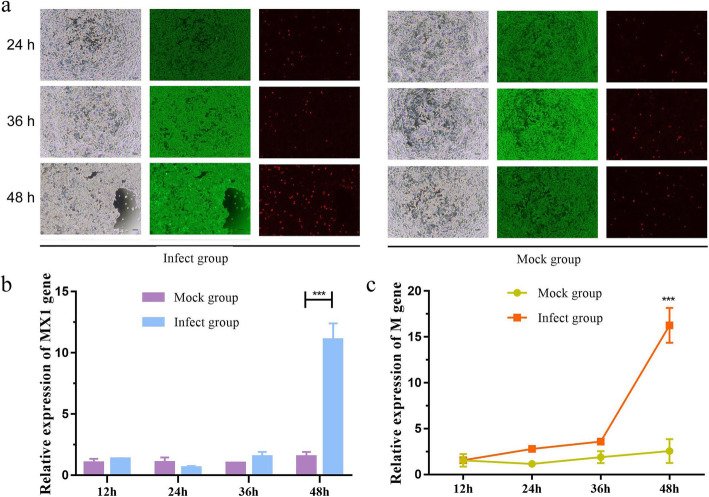
PEDV infection in IPEC-J2 cells. a Fluorescence-based cytotoxicity assay (using the LIVE/DEAD Viability/Cytotoxicity kit) of IPEC-J2 cells treated with PEDV strain CV777 at different time points. The green color indicates live cells stained by Calcein-AM and the red color indicates dead cells stained by propidium iodide. The green color was predominant in all viability assays, with only a few red (dead) cells appearing randomly. A significantly higher percentage of apoptotic cells (live (green)/dead (red)) are observed in the infected groups than in the controls (magnification, ×40; bars, 200 μm). b The mRNA expression level of MX1 gene in PEDV-infected IPEC-J2 cells as measured by qRT-PCR at different time points. c The mRNA expression level of M gene in PEDV-infected IPEC-J2 cells as measured by qRT-PCR at different time points. Data derive from three independent experiments and were analyzed by one-way ANOVA. Data are presented as the mean ± SD (n = 6). *** represents p < 0.001

