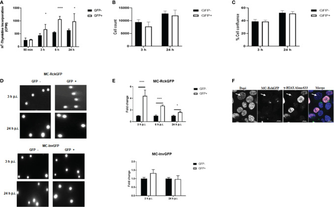
Figure 4.
Rck induces host cell DNA damage. HCT116 cells were infected with MC-RckGFP or MC-InvGFP strain. Infected cells (GFP+) and uninfected cells (GFP-) were sorted and then (A–C) re-cultivated in presence of H3-Thymidine. At the indicated times, the incorporation of H3-Thymidine was measured (counts per minute: cpm) (A), the number of cells per well (B) and the cell confluence per well were estimated (C). Data show the mean ± SD of one experiment (n = 5), representative of three experiments. (D, E) DNA damage assessment was performed using an alkaline comet assay. (D) Representative images of comets are shown in GFP− and GFP+ cells at the indicated times. (E) For each cell population, the tail extent moment (TEM) parameter was measured with the CometScore software. The data represent the mean ± SD of the fold change of the TEM studied parameters. At least 50 comets were analyzed on three slides/sample over three independent experiments. Data were compared using a t-test (**p < 0.01; ****p < 0.0001; GFP− vs. GFP+). (F) Expression and localization of γ-H2AX in HCT116 infected with MC-RckGFP. At 6 h p.i., infected cells underwent immunofluorescence staining using a mouse anti-γ-H2AX monoclonal antibody and an Alexa Fluor 633-conjugated secondary antibody (shown in red). Nuclei were stained with Dapi (shown in blue), and bacteria expressing GFP (shown in green) were visualized by fluorescence microscopy. A non-infected cell is indicated with a white arrow.

