Figure 7.

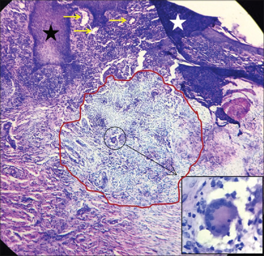
Histopathology of lupus vulgaris shows acanthosis (black star), hyperkeratosis (white star) and dilated capillaries (yellow arrows) and granuloma (red circle). Inset: Langhans giant cell. [H and E, 10×]

Histopathology of lupus vulgaris shows acanthosis (black star), hyperkeratosis (white star) and dilated capillaries (yellow arrows) and granuloma (red circle). Inset: Langhans giant cell. [H and E, 10×]