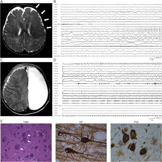
Figure 1. Patient 1: MRI, EEG, and anatomopathologic findings.
(A) Axial T2-weighted MRI section showing an extensive dysplastic area involving the entire left frontal lobe (arrows). (B) EEG recording at 6 months showing left frontocentral and anterior temporal ictal activity. (C) Axial T2-weighted MRI performed at 11 months after complete anatomic hemispherectomy. (D) EEG recorded at 15 months, showing build up of a right anterior frontal discharge. (E) Representative immunostaining of dysplastic brain tissue from the left frontal lobe. Hematoxylin-eosin (H&E) staining shows dysmorphic neurons (white arrows). Phosphorylated RPS6 (PS6) staining demonstrates hyperactivation of the mammalian target of rapamycin pathway. Antineurofilament (NF) staining demonstrates neurofilament protein accumulation in dysmorphic neurons. Scale bar = 50 μm.

