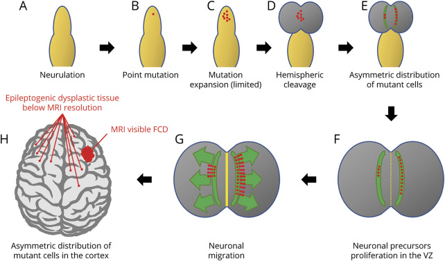
Figure 4. Schematic representation of the mechanisms that would lead to asymmetric distribution of mutant cells in the brain hemispheres.
After neurulation (A), a single point mutation occurs in a neuronal progenitor (B) and is passed to a limited number of daughter cells (C). At hemispheric cleavage (D), mutant cells migrate in the 2 hemispheres in different proportions because of their asymmetric initial distribution (E). After neuronal proliferation in the ventricular zone (VZ) (F), and neuronal migration (G), the asymmetric distribution of mutant cells in the 2 hemispheres causes a visible area of cortical dysplasia where the concentration of mutant cells is higher and only small foci of dysplastic tissue below MRI resolution where the percentage of mutant cells is very low (H). Note that even in the hemisphere harboring the main area of cortical dysplasia, mutant cells are present outside the limits of the visible abnormality and are the cause of seizure recurrence after surgery. FCD = focal cortical dysplasia.

