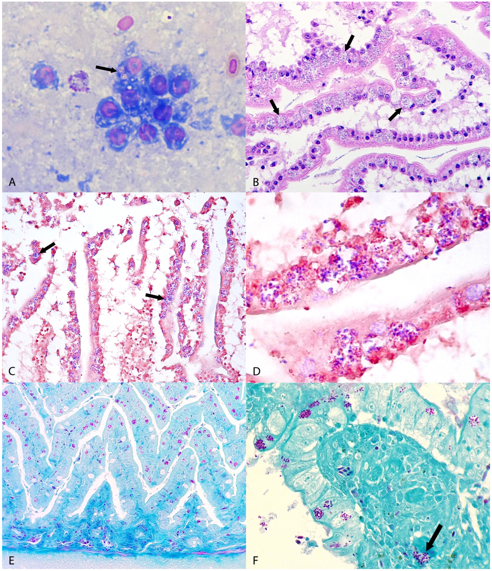
Fig. 1.

Microscopic findings from avian tissues. (A) Jejunum; canary, case No. 2. Oval to elliptical microorganisms with a clear cytoplasm and an eccentric nucleus compatible with microsporidian parasite are observed in the apical cytoplasm of enterocytes (arrows). Diff Quick. (B) Jejunum; European goldfinch, case No. 1. Note intense infection of the apical cytoplasm of enterocytes lining villi with microsporidial-like colonies located within parasitophorous vacuoles (arrows). HE. (C) Jejunum, European goldfinch, case No. 1. The microsporidial-like colonies in Fig. 1B stain gram positive (arrows). Gram. (D) Jejunum, European goldfinch, case No. 1. Higher magnification of gram positive microsporidial spores shown in Fig. 1C. Gram. (E) Jejunum, canary, case No. 2. Note abundant purple microsporidial colonies within the cytoplasm of enterocytes. Stamp. (F) Small intestine, lovebird, case No. 3. Note purple microsporidial colonies within the apical cytoplasm of enterocytes and in the lamina propria of villi (arrow). Stamp.
