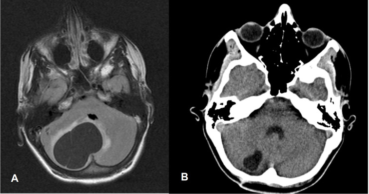
Figure 2.
(A) Aged 14 years. Preoperative T2-weighted-Fluid-Attenuated Inversion Recovery (FLAIR T2) MRI image of the brain showing right-sided cerebellar cystic tumour (cerebellar haemangioblastoma) with slight peritumoral oedema and a maximum cross-section of 56 mm, leading to compression of the fourth ventricle and slight displacement to the left (enhancing mural nodule and supratentorial obstructive hydrocephalus not visible). (B) Aged 16 years. CT scan image of the brain showing small residual cyst only with cross-section of 20 mm without any compression of the fourth ventricle.

