Figure 1.
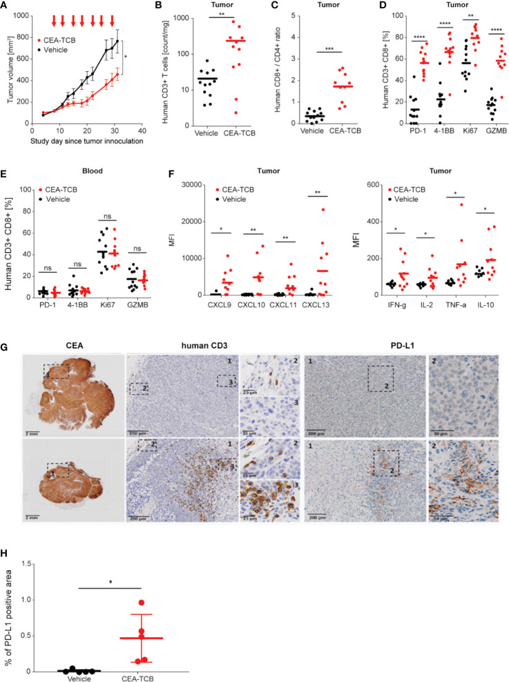
Treatment with CEA-TCB induces tumor growth inhibition and leads to increased frequency of tumor-infiltrating human T-cells and a tumor-specific T-cell activation in MKN-45-bearing hematopoietic stem cell humanized mice. Hematopoietic stem cell humanized NOG mice were inoculated subcutaneously with 1 × 10⁶ MKN-45 cells and treated with either buffer (vehicle; n=12) or with 2.5 mg/kg i.v. of CEA-TCB (n=12) twice weekly starting with a tumor volume of ~150 mm3 (Day 8). At termination (Day 32), blood and tumors were harvested for subsequent flow cytometry, histological and cytokine analysis (ImmunoPD data). (A) Tumor growth kinetics revealed a tumor growth inhibition (TGI) of 62%. Arrows indicate treatments (seven in total). (B–E) Flow cytometry analysis of tumor and blood in vehicle- and CEA-TCB-treated animals showing the frequency of tumor-infiltrating T-cells (B) and ratio of CD8+ to CD4+ T-cells in the tumor tissue (C), the expression of activation markers in tumor (D) and blood (E). (F) Cytokine/chemokine expression in tumor lysates. (G) Representative histological staining for human CEA, CD3, and PD-L1 on paraformaldehyde fixed tumor samples from vehicle (upper row) and CEA-TCB-treated animals (lower row). (H) Quantification of PD-L1 staining by IHC. (A) Data are mean ± SEM; (B–F, H) solid bars represent mean values; p-values are two-tailed unpaired t-test; *p < 0.05; **p < 0.01; ***p < 0.001; ****p<0.0001.

