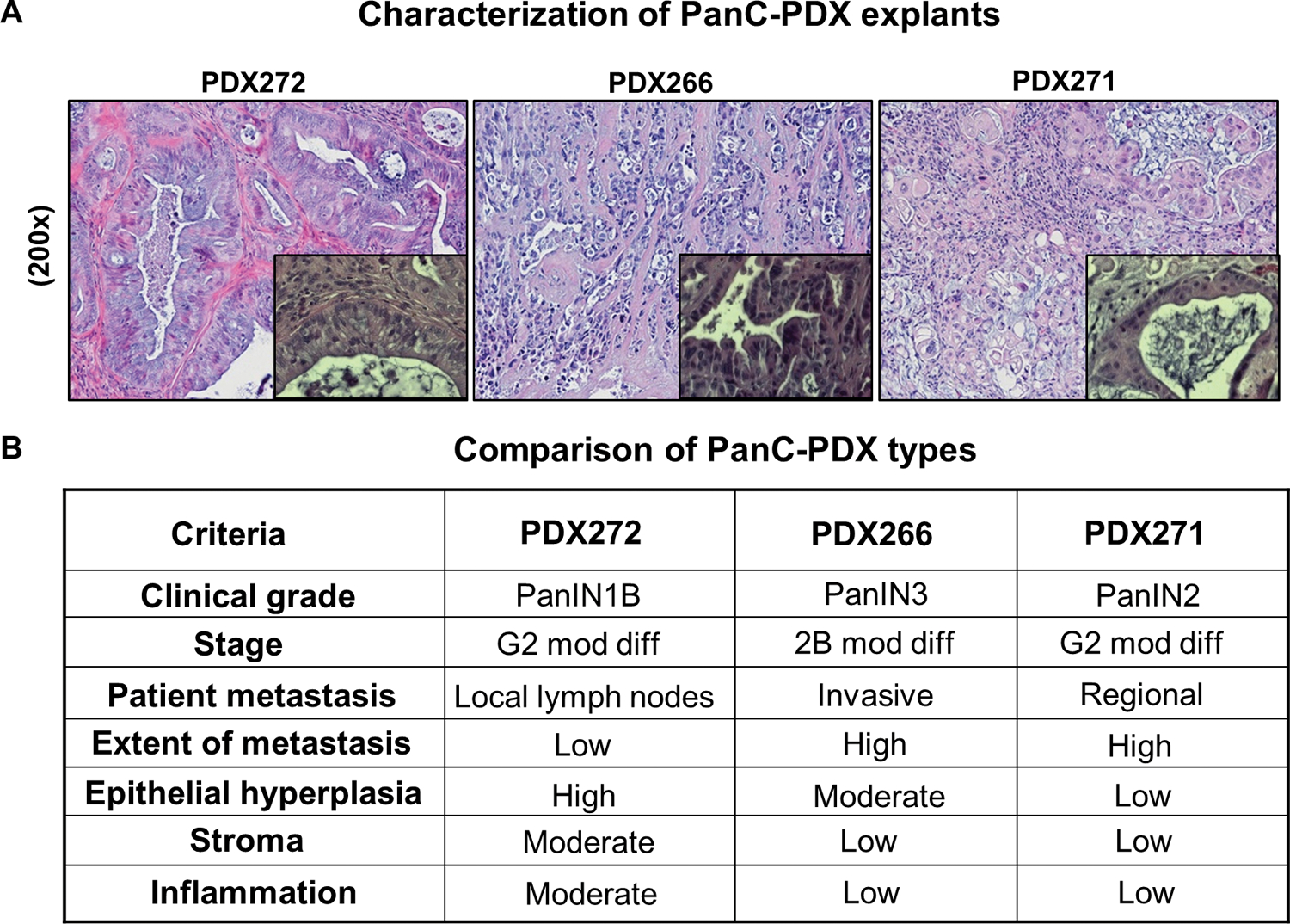
Figure 2. Comparative histopathological characterization of PanC-PDX272, PDX266, and PDX271 explants.

For our PDX study, excess tumor tissues not needed for diagnosis from Whipple resection of human pancreatic tumor specimens (patients without preoperative chemo- or radiation therapy) were used to propagate the PDX-explants in vivo in the PanC-PDX bank (UC Denver-AMC). (A) Representative pictographs (x200) of hematoxylin and eosin (H&E) stained PDX explants; magnified insets depicted at x400 magnification highlight the progression/clinical stage of PanC as seen by the ductal morphology. (B) A summary of the histological features of these explants provides details on PanC grade/stage, extent of metastasis, inflammation, epithelial hyperplasia, and stromal pattern. Moderately differentiated adenocarcinoma (mod diff).
