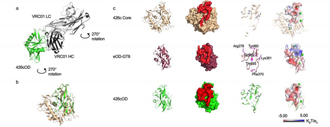
Figure 2. Structures of three VRC01 germline-targeting immunogens, related to supplemental tables 2-4.
(a) Crystal structure of 426cOD (green) bound to human mature VRC01 Fab (light chain in gray, heavy chain in black) shown in cartoon representation. (b) Overlay of 426c Core (tan, PDB ID 5FA2) and 426c OD (green). (c) Structural comparison of the three germline-targeting immunogens. From left to right: Cartoon representation. Surface representation of immunogens with glVRC01 epitope highlighted in red. glVRC01 epitopes shown in stick representation: eOD-GT8 residues that differ from those on 426c Core and 426cOD are highlighted in magenta. Electrostatic potential of glVRC01 epitopes. The location of potential NLGS around the VRC01 epitope on the three antigens are indicated by green dots (electrostatic potentials do not consider the glycans as exact glycosylation at these positions is unknown).

