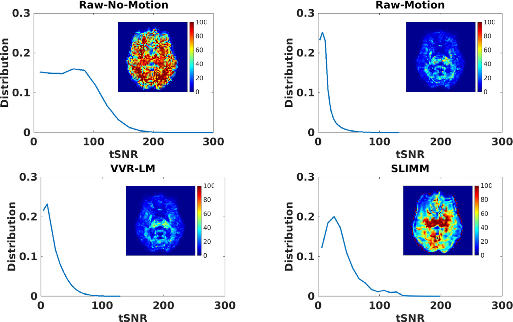
Fig. 4.

Distributions of the tSNRs over data from all subjects calculated by four methods compared to Raw-No-Motion and Raw-Motion data acquired with no motion and in-scanner motion respectively. The tSNR map calculated from the reconstructed fMRI time series by each method is also shown for an axial slice of a representative subject in each subfigure. These results show that our SVR method substantially improved the tSNR of the Raw-Motion data and outperformed VVR-LM. While tSNR is lost naturally due to motion in fMRI, the SVR methods were able to recover a large portion of tSNR despite the continuous subject movements that occurred during these scans. Our method, SLIMM, generated the best results according to the average tSNR values reported in Table 4.
