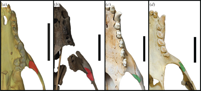
Figure 12.
Origin of masseter muscle. Ventral view of the left tooth row and zygomatic arch of Lycaon pictus SMNS 4461 (surface scan) (a), S. magnus USNM PAL 475486 (b), H. leptonyx NMV C27418 (c) and M. monachus NHMUK 1894.7.27.2 (d). Red patch indicates enlarged masseter origin that enlarges and interrupts the zygomatic arch profile. Green patch indicates reduced origin of masseter. Scale bars, 5 cm.

