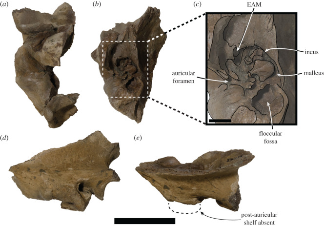
Figure 5.
USNM PAL 534034 right squamosal. (a) Ventral view and (b) medial view. (c) Close-up illustrated medial view of area designated by box in (b). (d) Lateral view. (e) Dorsal view, large dashed lines indicate regular monachine post-auricular shelf. EAM = external auditory meatus. Scale bar, 5 cm (a,b,d,e), and 1 cm (c).

