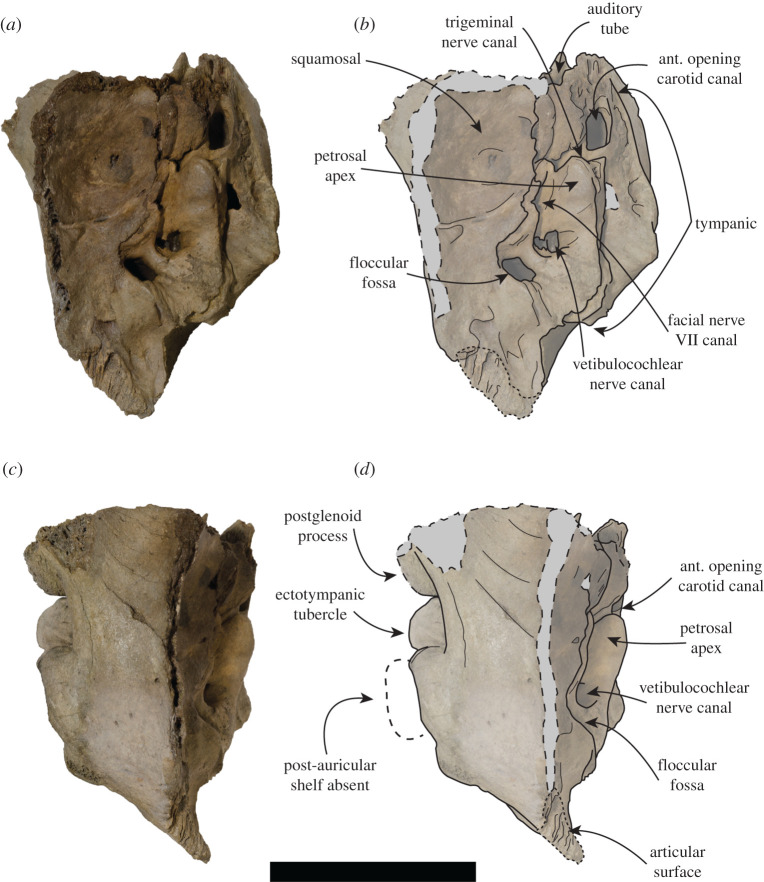
Figure 8.
USNM PAL 181601 left ear region. Photos (a,c) and drawings (b,d) of the left ear region. (a,b) Medial view and (c,d) dorsal view. Large dashed lines with grey areas indicate broken surfaces. Small dashed lines indicate articular surface. Large dashed lines with white area indicate regular monachine post-auricular shelf. ant = anterior. Scale bar, 5 cm.

